Northwestern celebrates 22 years as a top Fulbright producer
February 4, 2026
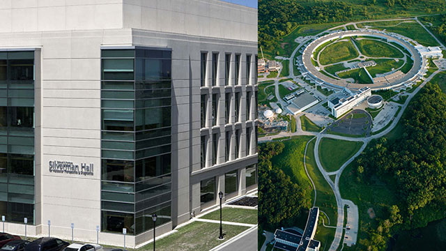
Northwestern University and its Chemistry of Life Processes Institute (CLP), the Advanced Photon Source (APS) at Argonne National Laboratory and several leading research universities are joining forces to launch a first-ever national hub for interdisciplinary, metallomics research and innovation.
The National Institutes of Health (NIH) has awarded Northwestern a five-year, $8.2 million grant to establish the Resource for Quantitative Elemental Mapping for the Life Sciences (QE-Map). The grant is from NIH’s Biomedical Technology Research Resources (P41) program.
QE-Map will enable unprecedented insights into the role of metals and other inorganic (non-living) elements in human health and disease.

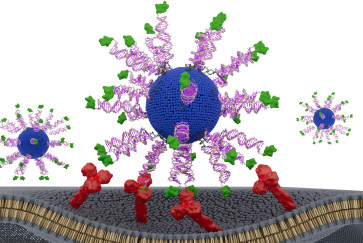
QE-Map will leverage Northwestern’s expertise in the areas of biophotonics, cell biology, inorganic chemistry and X-ray physics to pioneer cutting-edge imaging and detection technologies to deepen understanding of the interplay between metals and biological processes.
“Metal fluctuations play a critical regulatory role in a large number of biological processes,” said O’Halloran, the founding director of CLP and director of Northwestern’s Quantitative Bioelement Imaging Center (QBIC).

The resource will support 75 faculty research programs across Weinberg, McCormick and Northwestern University Feinberg School of Medicine as well as collaborators from Argonne, Howard Hughes Medical Institute’s Janelia Research Center, Georgia Tech, Johns Hopkins, Massachusetts Institute of Technology, University of California, Berkeley, University of South Carolina and University of Texas at Austin.
Investigators will provide the testbeds for new imaging technologies, tools, software and quantitative methods for metal analysis and localization. These new approaches will be applied to pioneering research in cardiac disease, fertilization and development, neurodegenerative diseases and metabolic diseases.
“The ability to map metals and metal dynamics in the brain opens important new avenues in neuroscience research, for neurodevelopmental and neurodegenerative diseases but also for the fundamental understanding of how neurons communicate,” said Yevgenia Kozorovitskiy, assistant professor of neurobiology at Northwestern.
“The field has focused on calcium signaling in neurons for a long time, but zinc and other metals serve critical synaptic signaling roles in neurons that remain poorly understood in addition to their functions as co-factors for important neuronal proteins,” she said.
 Quantitative elemental maps of the mouse brain obtained via LA-ICP-TOF-MS: Green regions correspond to high concentrations of zinc, especially prevalent in the hippocampal formation. Blue regions correspond to copper enrichment in periventricular zones, and red areas indicate iron abundance. (Sample courtesy of the Kozorovitskiy and O’Halloran groups, analysis software from Tofwerk AG).
A new mass spectrometry tool operated by QBIC will enable rapid 2D and 3D imaging of cells in tissue slices. Advances will include development of a completely integrated system with real-time multi-element imaging capabilities, standardized procedures for handling cryogenically preserved biological tissues and a universal cryo sample mounting system that can move between several different technologies that map and analyze elemental distributions.
The resource will also employ large sample format Scanning X-ray Fluorescence Microscopy (SXFM) utilizing one of the nation’s brightest synchrotron radiation sources: the Advanced Photon Source, a U.S. Department of Energy Office of Science user facility located at Argonne National Laboratory. The SXFM will provide researchers with organ-wide information on metal distribution.
Jacobsen, an expert in X-ray microscopy and trace metal quantitation, will lead efforts to increase the speed, cost-effectiveness and imaging sensitivity of SXFM and standardize approaches for cryogenically preserving biological tissues for universal adoption across platforms.
“We want to push further the boundaries of what can be done with X-ray imaging and apply it to biological systems,” said Jacobsen, a member of the Chemistry of Life Processes Institute. “The APS at Argonne is a great way to excite that X-ray florescence with a much higher rate and much higher spatial resolution than ever could be done in a laboratory system.”
A third resource technology, Photo Acoustic Spectroscopy (PAM), is pioneered by Zhang and Sun. PAM deploys a highly novel, modular ultrasonic detector that combines a unique chemical process to image both frozen samples and live tissue at unprecedented resolution. This approach works by scanning a laser across a sample, causing heat and color intensity to increase and triggering a shockwave that can be detected acoustically. Cheng and Sun will work with collaborators to engineer molecular probes that cater to specific experimental needs. These advances will provide better ways to image time-dependent fluctuations in the concentrations of specific metal ions.
“It’s almost like hearing the sound of a color,” O’Halloran said. “There are disorders of the human brain where sensory inputs get confused between them, but in this case, we’re actually using the absorbance of a photon of a certain wavelength of light, which gives rise to the observation of a color, creating a sound wave that will help us localize an image in the material. By listening to the photo acoustic detectors, we can create a map from the sound pattern. It’s an incredible idea that has never been shown before.”
QE-Map will occupy a unique place among the 40 NIH-funded national resources as the only one advancing technology for studying the role of metals in health and disease.
“We will be collaborating with dozens of biomedical research teams from around country with the goal of understanding the molecular mechanisms at work in a variety of metal-linked diseases, from Wilson disease, Menkes disease and Alzheimer’s, to Parkinson’s, heart disease and diabetes,” O’Halloran said. “Each of the investigative teams around the country have a particular question in which they need to know how much metal is in their sample and what is it doing to the sample. We can help answer all those questions.”