Northwestern celebrates 22 years as a top Fulbright producer
February 4, 2026

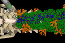
Northwestern University scientists have developed the most advanced organoid model for human spinal cord injury to date.
In a new study, the research team used lab-grown human spinal cord organoids — miniature organs derived from stem cells — to model different types of spinal cord injuries and test a promising new regenerative therapy.
For the first time, the scientists demonstrated that human spinal cord organoids can accurately mimic the key effects of spinal cord injury, including cell death, inflammation and glial scarring, a dense mass of scar tissue that creates a physical and chemical barrier to nerve regeneration.
When treated with “dancing molecules” — a new therapy that reversed paralysis and repaired tissues in a previous animal study — the injured organoids showed significant outgrowth of neurites, the long extensions of neurons that connect the cells to one another. The glial scar-like tissues of treated injured organoids also significantly diminished. These results give researchers further hope that the treatment, which recently earned an Orphan Drug Designation from the U.S. Food and Drug Administration (FDA), should improve outcomes for patients with spinal cord injuries.
The study was published today (Feb. 11) in the journal Nature Biomedical Engineering.
“One of the most exciting aspects of organoids is that we can use them to test new therapies in human tissue," said Northwestern’s Samuel I. Stupp, the study’s senior author and inventor of dancing molecules. “Short of a clinical trial, it’s the only way you can achieve this objective. We decided to develop two different injury models in a human spinal cord organoid and test our therapy to see if the results resembled what we previously saw in the animal model. After applying our therapy, the glial scar faded significantly to become barely detectable, and we saw neurites growing, resembling the axon regeneration we saw in animals. This is validation that our therapy has a good chance of working in humans.”
A pioneer in self-assembling materials and regenerative medicine, Stupp is the Board of Trustees Professor of Materials Science and Engineering, Chemistry, Medicine and Biomedical Engineering at Northwestern, where he has appointments in the McCormick School of Engineering, Weinberg College of Arts and Sciences and Feinberg School of Medicine. He also directs the Center for Regenerative Nanomedicine (CRN). Nozomu Takata, a research assistant professor of medicine at Feinberg and member of CRN, is the paper’s first author.
Grown in the lab from induced pluripotent stem cells, organoids are miniature, simplified versions of human organs. Although they are only partial organs, organoids mimic the tissue structure, cellular complexity and function of the real thing. This sophisticated mimicry makes organoids ideal for modeling human diseases, testing therapeutics and understanding organ development. Compared to testing treatments in animals and humans, testing in organoids is faster and much less expensive.
While other researchers have developed human organoids to investigate physiological aspects of the spinal cord, Stupp’s model represents a giant leap forward to find treatments for devastating, paralyzing human injuries. Measuring several millimeters in diameter, the organoids were large and mature enough to develop the injury model.
Stupp’s team grew the spinal cord organoids from stem cells over the course of months, allowing them to develop complex features including neurons and astrocytes. The team also was the first to add microglia — immune cells in the central nervous system — to simulate inflammatory responses to traumatic spinal cord injury.
“It’s kind of a pseudo-organ,” Stupp said. “We were the first to introduce microglia into a human spinal cord organoid, so that was a huge accomplishment. It means that our organoid has all the chemicals that the resident immune system produces in response to an injury. That makes it a more realistic, accurate model of spinal cord injury.”
After developing a mature spinal cord organoid, Stupp and his team wanted to examine the effects of injuries and subsequent treatment. First introduced in 2021, the dancing molecules therapy harnesses molecular motion to reverse paralysis and repair tissues after traumatic spinal cord injuries. It is part of the Stupp laboratory’s platform of supramolecular therapeutic peptides (STPs), technologies that use large assemblies of 100,000 or more molecules to activate cell receptors using the body’s own natural signals to regenerate and repair. (The concept of supramolecular therapies also is used in current GLP-1 drugs for weight loss and diabetes, an area that Stupp’s lab investigated nearly 15 years ago.)
Injected as a liquid, the dancing molecules therapy immediately gels into a complex network of nanofibers that mimic the extracellular matrix of the spinal cord. By fine-tuning the collective motion, or “dancing,” of the molecules within the nanofibers, Stupp’s team found the therapy connects more effectively with constantly moving cellular receptors.
“Given that cells themselves and their receptors are in constant motion, you can imagine that molecules moving more rapidly would encounter these receptors more often,” Stupp said in 2021. “If the molecules are sluggish and not as ‘social,’ they may never come into contact with the cells.”
In animal studies, a one-time injection administered 24 hours after severe injury helped mice regain the ability to walk in just four weeks. Compared to injections with slower-moving molecules, formulations with enhanced molecular motion had greater therapeutic efficacy, indicating increased bioactivity and cellular signaling.
To model spinal cord injury, Stupp’s team induced two types of common injuries. The researchers cut some of the organoids with a scalpel to simulate a laceration, like a surgical wound. For other organoids, the researchers applied a compressive contusion injury to simulate wounds that might occur in a serious car accident or from a steep fall.
Both injuries caused cells to die and a glial scar to form — just like in a real spinal cord injury.
“We could distinguish between the astrocytes that are a part of normal tissue and the astrocytes in the glial scar, which are large and very densely packed,” Stupp said. “We also detected the production of chondroitin sulfate proteoglycans, which are molecules in the nervous system that respond to injury and disease.”
After simulating injuries, Stupp’s team tested the effectiveness of the dancing molecules. When applied to the injured organoids, the liquid therapy gelled to form a scaffold. The therapy calmed inflammation, reduced glial scarring, caused neurites to extend and encouraged neurons to grow in neat, organized patterns.
A type of neurite, called an axon, is often severed during spinal cord injury, disconnecting the communication network among neurons. That disconnection results in paralysis and a loss of sensation below the injury site. Regenerating neurites could reestablish these connections to prevent or reverse these devastating outcomes.
Stupp attributes the treatment’s success to its supramolecular motion — or the ability of the molecules to move rapidly or even temporarily leap out of the nanofibers. Testing the therapy on healthy organoids only confirmed Stupp’s hunch.
“Before we even developed the injury model, we tested the therapy on a healthy organoid,” he said. “The dancing molecules spun out all these long neurites on the surface of the organoid but, when we used molecules that had less or no motion, we saw nothing. This difference was very vivid.”
Next, Stupp’s team plans to build even more advanced organoids to further refine their model. They also plan to develop a human spinal cord organoid that models older, chronic injuries, which typically have more stubborn scar tissue. With further work, Stupp said his group’s mini spinal cords also could be used in personalized medicine, by creating implantable tissue using a patient’s own stem cells to avoid immune rejection.
The study, “Injury and therapy in the human spinal cord organoid,” was supported by the Center for Regenerative Nanomedicine at Northwestern University and a gift from the John Potocsnak Family for spinal cord injury research.