
Embed Video:
<iframe width="560" height="315" src="https://www.youtube.com/embed/P3uSHn63nrk" frameborder="0" allow="accelerometer; autoplay; encrypted-media; gyroscope; picture-in-picture" allowfullscreen></iframe>
New technology illuminates the messages that cells leave behind
EMBARGOED UNTIL 5 A.M. EST (U.S.) ON TUESDAY, NOVEMBER 18, 2025
EVANSTON, Ill. --- In stunning new time-lapse videos, biological nanoparticles scoot and flit across a starry field of glowing dots. Guided by the invisible chemistry of attraction, these microscopic travelers eventually group together to form perfectly round, glowing circles on a black surface.
These mesmerizing new videos are made possible with LEVA (light-induced extracellular vesicle and particle adsorption), a groundbreaking new technology from Northwestern University and The Ohio State University.
LEVA is the first tool that enables scientists to precisely arrange tiny biological packages called surface-bound extracellular vesicles and particles (EVPs). Cells release these tiny packages into biofluids and leave them behind in tissues, where they help pass messages to other cells. By signaling cells to move or to repair damage, EVPs appear to influence many important processes in the human body, including wound healing, infection, regeneration and cancer spread.
Using LEVA, scientists can watch these tiny biological couriers as they interact with cells in real time. Then, researchers can probe how EVPs’ messages accelerate healing, help defend the body or proliferate disease.
The study will be published tomorrow (Nov. 18) in the journal Nature Methods. It marks the first rapid, scalable, high-resolution tool for controlling EVPs without using antibodies, chemical tags or capture molecules.
“Our research provides scientists with a powerful new tool to understand how cells communicate through the ‘breadcrumb trails’ they leave behind during movement in both healthy and disease contexts,” said Northwestern’s Colin Hisey, who co-led the work. “A better understanding of their role could lead to new treatments for diseases and improved wound healing therapies. The technique’s versatility means it can be adopted by researchers worldwide to accelerate discoveries in multiple areas of human health.”
Hisey is an assistant professor of biomedical engineering at Northwestern’s McCormick School of Engineering. He co-led the work with Xilal Y. Rima and Jacob Doon-Ralls, a postdoctoral fellow and graduate fellow, respectively, at The Ohio State University. The study’s senior author is Eduardo Reátegui, an associate professor of chemical and biomolecular engineering at The Ohio State University.
As cells move through the body, they naturally release tiny membrane-wrapped EVPs, which carry proteins, RNA and other molecular cargo. Once released, EVPs act as messengers among cells to shape various biological processes, including immune responses and tissue growth.
Traditionally, scientists have studied EVPs suspended in liquid. But Hisey and his team wanted to explore what happens when EVPs are fixed in place, acting as a roadmap for traveling cells. LEVA works by shining ultraviolet light onto a tiny array of mirrors, and then onto a surface, to create a stencil-like pattern. Areas exposed to the light undergo a chemical change, becoming sticky to EVPs. Unexposed areas, however, remain neutral.
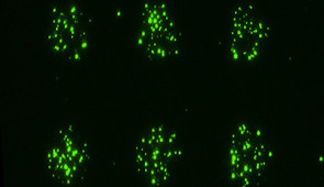
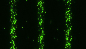
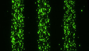
When EVPs are introduced, they naturally attach to the exposed regions, forming precise patterns such as dots, lines, gradients, trails or even complex images. This allows scientists to arrange EVPs into controlled shapes and trails that mimic how they might be arranged in human tissues.
“EVPs appear to play a crucial, but poorly understood, role in cancer migration and metastasis, wound healing and immune responses,” Hisey said. “Previously, scientists lacked the tools to study them quantitatively and systematically. LEVA uses controlled ultraviolet light to attract these vesicles with subcellular precision based on their innate properties. This wasn’t possible before, and thanks to our interdisciplinary team, this technology comes right at a time when this field is gaining a lot of attention and momentum.”
After developing patterns of EVPs, Hisey and his collaborators wanted to study how they interact with cells in real time. They used LEVA to create precise shapes of EVPs from bacteria, simulating an infection. Then, the team added isolated human neutrophils, a type of white blood cells that act as the immune system’s first responders.
Neutrophils rapidly detected and swarmed toward the patterned bacterial EVPs. Clustering tightly over EVPs, the neutrophils mimicked how they might gather at the site of a real wound or infection in the body. In accompanying time-lapse videos, viewers can watch the army of white blood cells wiggle and jiggle across the flat surface, covering the patterns made of bacterial EVPs in different shapes including dots and even stars.
The experiment shows that EVPs alone — without live bacteria — act as strong chemical beacons for immune cells. With LEVA, scientists can study this signal-response behavior with high precision, which could help researchers better understand immune signaling and inflammation.
“Neutrophils have evolved to recognize the antigens present on bacterial cells and, hence, also bacterial EVPs because they are so similar,” Hisey said. “Once neutrophils come into contact with and initially sense the EVPs, they undergo dynamic responses that we’re still trying to understand. This is something our platform can help us study.”
Next, Hisey and his team plan to expand LEVA beyond flat, transparent surfaces to more complex, three-dimensional and biologically relevant materials to better mimic the conditions inside the human body. By fine-tuning patterns and gradients, the team eventually hopes to decode the rules of EVP-driven cell behavior, from guiding tissue regeneration to intercepting cancer spread.
“We want to apply LEVA across multiple disease areas to systematically map how different types of surface-bound vesicles affect cell behavior in various conditions and configurations, with an initial focus on cancer metastasis, wound healing and immune responses to pathogenic EVPs,” Hisey said. “Our long-term goals include developing therapeutic strategies that harness or block these vesicle-mediated cellular communications and expanding the technique to study how nanoparticles interact with surfaces in a purely materials engineering context.”
The study, “Light-induced extracellular vesicle and particle adsorption,” was supported by the National Institutes of Health, Ohio State’s Center for Cancer Engineering, OK-PROS and LEGACY program, and the Burroughs Wellcome Fund.
<iframe width="560" height="315" src="https://www.youtube.com/embed/P3uSHn63nrk" frameborder="0" allow="accelerometer; autoplay; encrypted-media; gyroscope; picture-in-picture" allowfullscreen></iframe>
Please credit images to Colin Hisey/Eduardo Reategui